


Introduction to XRD Device:
X-ray diffraction is a non-destructive method with multiple applications, providing comprehensive information about chemical compositions and crystalline structures of natural and industrial materials. Each crystal has its unique X-ray pattern, which serves as a fingerprint for its identification. The most common use of XRD is identifying crystalline compounds based on their diffraction pattern. Other applications of XRD include geology, materials science, environmental science, chemistry, physics, pharmaceuticals, and more.
Introduction to XRF Device:
There are two main methods for measuring element content: chemical analysis, mainly by titration, and non-destructive analysis using X-rays, called XRF (X Ray Fluorescence). The advantages of XRF over chemical methods include speed, low cost, and acceptable accuracy. XRF measures the wavelength and intensity of fluorescent radiation emitted by atoms in a sample, providing both qualitative and quantitative elemental analysis. XRF has wide applications across many sciences and is now considered an essential instrument in research laboratories. Its high speed allows analysis of many elements quickly, and its non-chemical approach is cost-effective and environmentally friendly.
Key Services Provided in Mineralogy Laboratory:
- Analysis by XRD, obtaining spectra and diffraction patterns, determination of Miller indices, etc.
- Determining asbestos content in compounds
- Identification of phases or any type of mineral in rocks, soils, minerals, etc.
- Study of crystal structure
- Determining amorphous or crystalline nature of compounds
- Semi-quantitative phase percentage determination
- Determination of FWHM
- Nanoparticle size measurement via Debye-Scherrer method
- Chemical composition study of minerals
- Qualitative and quantitative elemental analysis
- Bulk analysis of main elements (Si, Ti, Al, Fe, Mn, Mg, Ca, Na, K, P)
- Analysis of rare and heavy elements (Ba, Ce, Co, Cr, Cu, Ga, La, Nb, Ni, Rb, Sc, Sr, Rh, U, V, Y, Zr, Zn)
- Results reporting in oxide form
- Capability to introduce 100 samples simultaneously to the device
For downloading XRF and XRD sample submission form, visit Sample Submission Form.
XRF Device Principle
X-ray radiation from the XRF tube hits atoms in the sample, producing fluorescent waves. Primary radiation excites electrons from various energy levels, leaving atoms in an unstable state. Electrons from higher energy levels then fill lower energy levels (less energy).
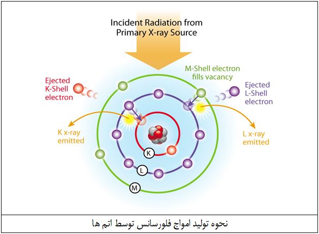
During these atomic transitions, excess energy is emitted as X-ray waves with specific wavelengths. Measuring these wavelengths identifies the element, with diffraction crystals helping to measure the emitted wavelengths.
Measuring the intensity allows determination of each element's abundance in the sample.
XRF Device Accuracy
The accuracy of element measurement depends on sample preparation and the standard sample used for device calibration.
Sample Preparation:
One of the most important parts of any laboratory is sample preparation. The goal is to obtain a representative and finely ground sample. The process includes:
1) Drying: All samples are completely dried at the required temperature.
2) Crushing: Samples are crushed by jaw crushers to sizes of about 3–7 mm.
3) Sampling: From the crushed sample, about 50 g of representative sample is selected using a riffle splitter.
4) Grinding: Prepared sample is ground by disc mill to about 75 μm (200 mesh).
5) Pellet preparation: Two types of pellets are made:
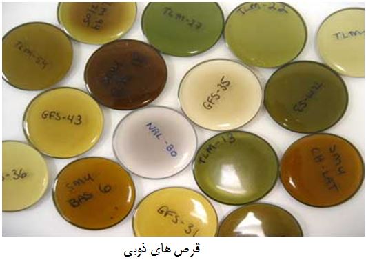
Fusion pellets:
In fusion pellets, the sample is melted using fluxes, mostly containing one boron element (especially lithium borate).
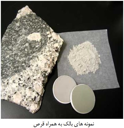
Pressed pellets:
In pressed pellets, sample powder is mixed with an inert binder and pressed into pellets for device analysis.
Fusion pellets
Finally, prepared pellets are placed in special cassettes for the device. Each sample analysis takes about 15 minutes, with data processing being the more important part, performed by skilled personnel.